by booker | Oct 9, 2020 | News
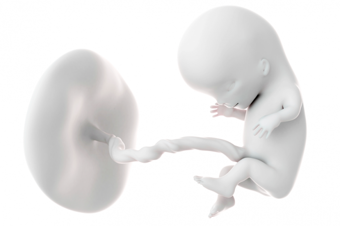
While initially the 11-13 weeks’ scan was performed with the sole purpose to measure the nuchal translucency and calculate the risk for Down syndrome, it evolved over the years to become a full anatomy check of the fetal organs. The completeness of the examination...

by booker | Oct 9, 2020 | News
The dating of the pregnancy and thus the calculation of the expected date of delivery (EDD or the due date) is based on the some prerequisites such as: the mother has a regular 28-day cycle, she is sure of her last period, she has not recently taken contraceptive...

by booker | Oct 9, 2020 | Consulting
In some cases serious problems occur in the mother and/or the fetus or there is a family history of medical conditions. The diagnosis and the management of these pregnancies often requires the collaboration of a team of doctors, each one contributing to solving their...

by booker | Oct 3, 2020 | Ultrasounds
This is the third detailed scan during the pregnancy carried out usually between 28 and 34 weeks. In special circumstances it may need to be repeated according to the mother’s and the fetus’ condition. The aim of the examination is to check the placenta, the amniotic...
by booker | Oct 2, 2020 | Ultrasounds
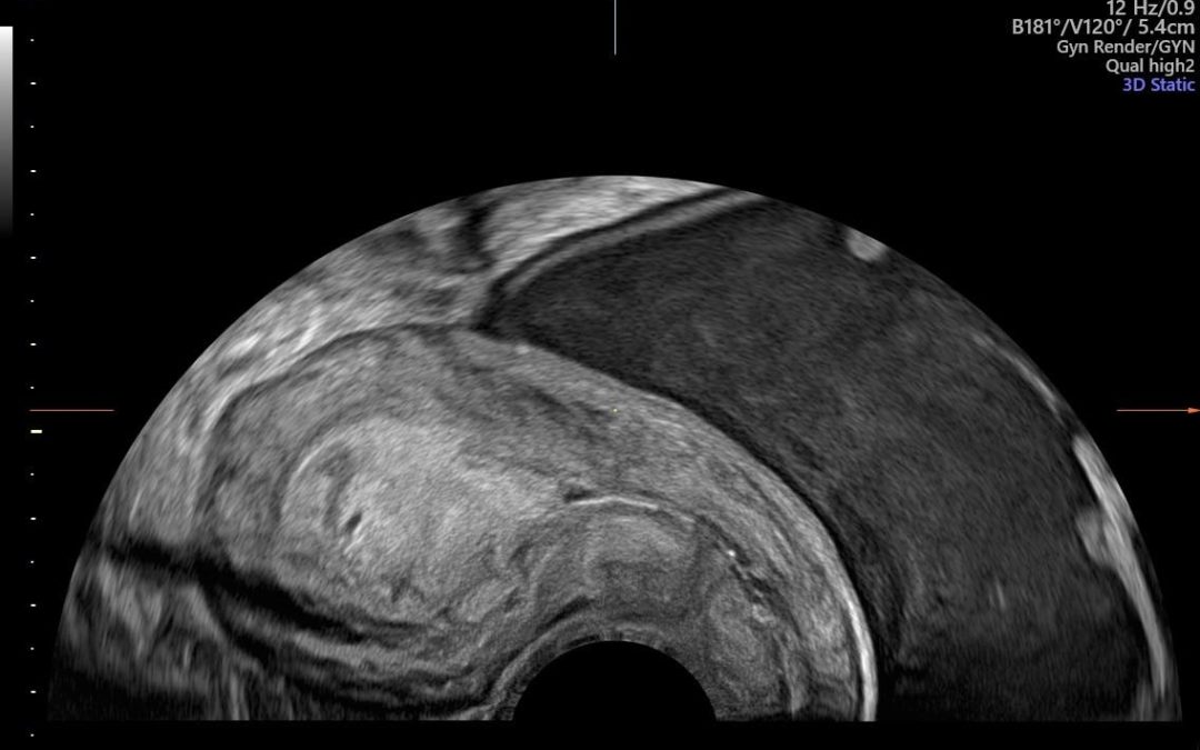
The ultrasound examination of the womb and the ovaries is nowadays an integral part of the gynecological examination. The visualization of the internal female organs can reveal conditions as yet unsuspected (such as fibroids) or provide an explanation for existing...